The EpiOcularTM Model
주문은 최소 1개월 전에 하셔야 합니다.

Features
EpiOcular Tissue Model
- Cornea-Like 3-D Tissue Structure
- Alternative to Draize Rabbit Eye Test
- Excellent correlation - In Vivo to In Vitro Test Results
- "Sub-Draize" (Mild, Milder, Mildest) Product Testing Possible
- Topical Testing of Undiluted and Water Insoluble Materials Possible
- 15 YEARS of Publicly Available QC Data
- Also Available in 24 and 96-Well Formats
- Extensive Number of Materials, Products Tested
General MatTek Tissue Features
- Unsurpassed Long-Term Tissue Reproducibility
- Lot-to-Lot, Year-to-Year
- 3-Dimensional, Highly Differentiated Tissue
- Metabolically, Mitotically Active
- Produced from Normal (Non-Transformed) Human Cells - Ideal for Genomics Studies
- Produced in Easily Handled Cell Culture Inserts
- Grown in Completely Serum-Free Media System
- Quantifiable, Objective Test Endpoints
- Cost Effective Alternative to Animal and Human Clinical Testing
Applications
EpiOcular Tissue Model
- Ocular Irritation Determination
- “Sub-Draize” Mild, Milder, Mildest Ocular Irritation Testing
- Draize Score Determination for Surfactants and Surfactant-based Products
- Cosmetics, Consumer Products, etc.
Data Sheet
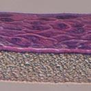
MatTek's EpiOcular™ corneal model consists of normal, human-derived epidermal keratinocytes which have been cultured to form a stratified, squamous epithelium similar to that found in the cornea. The epidermal cells, which are cultured on specially prepared cell culture inserts using serum free medium, differentiate to form a multi-layered structure which closely parallels the corneal epithelium.
HISTOLOGY:
EpiOcular vs. Rabbit Cornea
Formalin Fixed, Paraffin Embedded, H&E Stained
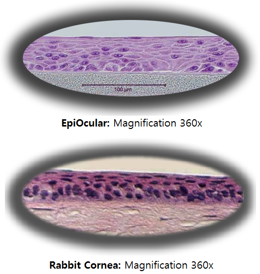
The EpiOcular tissue model exhibits in vivo-like morphological and growth characteristics which are uniform and highly reproducible. EpiOcular consists of highly organized basal cells which progressively flatten out as the apical surface of the tissue is approached, analogous to the normal in vivo corneal epithelium. EpiOcular is mitotically and metabolically active and releases many of the pro-inflammatory agents (cytokines) known to be important in ocular irritation and inflammation.
The protocols for using the EpiOcular System are clear and straightforward. EpiOcular has been utilized with several common tests of cytotoxicity and irritancy, including MTT, IL-1α, PGE2, LDH, and sodium fluorescein permeability. Technicians find EpiOcular's rigid substrate design easy to handle in routine repetitive testing environments and scientists find that they are able to perform discriminating tests due to the model's high level of reproducibility. In fact, "Sub-Draize" testing is possible with EpiOcular, allowing quantifiable discrimination among mild, milder, and mildest product formulations.
Various industrial and toxicology laboratories are actively seeking alternatives to whole animal testing. Cosmetic, household product, pharmaceutical and petrochemical companies have initiated in vitro toxicology testing to evaluate their raw materials and final product formulations. A growing body of data indicates that EpiOcular effectively provides a non-animal means to assess ocular irritation and other toxicological issues.
For all of these reasons, the EpiOcular model provides a predictive, morphologically relevant in vitro means to assess ocular irritancy.
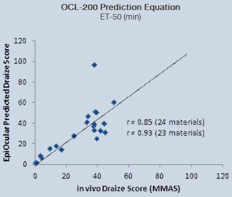
Figure 1: Determination of EpiOcular prediction model - Correlation of EpiOcular MTT ET-50 results to Draize rabbit eye data.
|
|
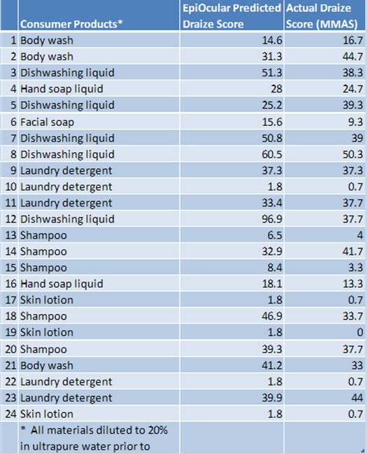
All materials diluted to 20% in ultrapure water prior to testing (density >0.95g/ml)
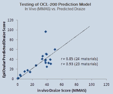
Figure 2: Comparison of actual Draize to predicted Draize score based on correlation equation (Figure 1).
|


'Human tissue' 카테고리의 다른 글
| The EpiAirway™ Tissue Model (MatTek) (0) | 2013.06.29 |
|---|---|
| The EpiVaginal ™ Tissue Model (MatTek) (0) | 2013.06.29 |
| The Melanoma Skin Model (MatTek) (0) | 2013.06.29 |
| The MelanoDerm ™ Skin Model (MatTek) (0) | 2013.06.29 |
| The EpiOral ™ Tissue Model (MatTek) (0) | 2013.06.29 |